
|
Casuistics: Early Thermocoagulation instead of Surgery to cure Keratoacanthomas
|
|
|
Casuistics: Early Thermocoagulation instead of Surgery to cure Keratoacanthomas
|
 < health
< back
< health
< back
Summary: Keratoakanthomas are fast growing tumors exhibiting pressure pain. They can initially be confounded with early furunculosis, may double in size within a few days but have a very low or absent tendency to metastasize. Surgical removal with 4-5mm healthy surrounding tissue avoids relapses but may cause problems for wound covering especially for ear or nose. Insensitivity against locally administered antibiotics discriminates them early on (4-6 days) from infectious lesions with pus formation. During this time they can be removed without relapse or scar formation by a series of pin point thermocoagulations.
Painful nodule on the right cheek: A painful pinhead sized nodule formed in June 2018 around 2cm below the zygomatic arc. It was treated with an antibiotic ointment (Aknemycin, 20mg Erythromycin/g) assuming the development of a small furuncle. Three day appklication of the ointment did not influence the growth of the nodule and no pus was formed. The lesion was approximately 1mm high and considered for a short time as a fast growing wart (fig.1). The nodule was deeply treated on day 11 with 30% trichloracetic acid including 2mm into the healthy tissue around. This destroyed the center of the lesion but not its border region. Actikerall (5mg/g 5-Fluoruracil, 100mg/g Salizylsäure) was subsequently applied on the tumor and 2mm around to reduce cell growth. This removed the surface layers of the tumor but did not stop its further extension.
Therapy: After 6 weeks a biopsy was taken in a university dermatolgy hospital. The tissue was identified as squamous cell carcinoma with Keratoacanthoma as differential diagnosis and surgical excision was recommended. The distinction beteen squamous cell carcinoma and keratoacanthoma by morphological criteria is difficult (1) in contrast to the comparatively clear discrimination by molecular biological analysis (2). Squamous cell carcinomas develop rather slowly without pain, while Keratoacanthomas grow fast and show pressure pain. A second opinion was sought urging surgical excision, The excision was performed on day 66 after perception of the first pain at tumor sizes of 14 and 15mm on days 28 and 62. Tumour growth had become slower during during this period, possibly due to the earlier treatment. The tumor was successfully removed without relapse and identified again as squamous cell carcinoma with differential diagnosis keratoacanthoma although its clinical development ressembled more to a keratoacanthoma.


Fig.1 Keratoacanthoma of the right cheek on days 7 (left), 28 (center left), 62 (center right) as well as the several cm long scar 48months after surgical removal on 66.Tag (right) (click on images for enlargement).
Painful right nasal wing: A painful and reddened small area developed in January 2021 on the right nasal wing. Aknemycin treatment did not improve the situation, the lesion continued growing, no pus was formed and the lesion measured around 4mm in diameter by day 7 (fig.2 top left), suggesting a second keratoacanthoma. Thinking of the earlier experience the tumor with grow until surgical removal to a significant size. Cutting it out around 4-5mm in the healthy surroundng tissue would generate an uncoverable defect, requiring a partial nose prothesis. So faster tumor removal by thermocoagluation was required.
Burn out with heated nail head: A nail with a nailhead corresponding to the tumor size was heated with a gas flame until red heat on day 7 and tipped several times gently onto the tumor surface, considering that the tumor was superficially growing above the skin basal membrane. The first touch was quite painful, but thermodestruction of superficial nerve endings led to a certain local anesthesia effect, facilitating subsequent touches. The wound was treated on day 11 with 3% hydrohen peroxide (H2O2) solution to remove the eschar as well as dead tissue- It was clean and seemed tumor free (fig.2 top, center left) but an illusion upon wound cleaning on day 19 (fig.2 top, center right). The tumor was hit in the center with significant proliferation in the border areas, icluding the danger of tumor growth into the nose interior where it would be unreachable for thermocoagulation.
Electronic soldering iron (40Watt): A soldering iron with a noncorroding V2A steeltip (2x2 mm2) at 340C was used in a second thermocoagulation effort. Leaning the ellbow against the bathroom mirror permitted jitterfree, precise and careful touches of the tumor surface by the hot steel tip. 30% trichloroaceetic acid was applied onto the tumor and 2mm around in the healthy tissue to prophylactically coagulate tumor front cells. Cured areas did not show sharp pain upon careful fingertip touching. The lower tumor areas as well as the lateral tumor front towards the nose interior required numerous steel tip touchings. They were always applied with low pressure (fig.2 top right) to avoid unnecessary tissue loss in view of subsequent healing with as little as possible scar formation. This was successful as shown by a practically scar free end result (fig.2, bottom center & right) with total recovery of surface sensibility.


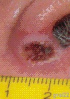


Fig.2 Painful swelling of the right nasal wing (7 days, top left). The tumor seemed to be removed by the immediate thermocoagulation, considering the clean wound surface on day 11 (top, center left) but a significant relapse was noticed on day 18 (top center right). The tumor center was well hit but proliferation occurred in the border areas. Slight repeated touches with the V2A steel tip of an electronic soldering iron (40W, 340C) on day 19 evaporated the tumor tissue (top right) with progressive wound healing on day 39 (bottom left) and day 69 (bottom center) until a practically scar free end result after 21 months (bottom right)(click on images for enlargement).
Requirement for early Intervention: Despite the satisfying result of the keratoacanthoma therapy on the right nasal wing (fig.2 bottom right), thermocoagulation should start earlier than 7 days (fig.3) when their development is still in the pinhead nodule size being at that time in contrast to the diffuse swelling and pus formation of infectious lesions.


Fig.3 A keratoacanthoma of the forehead center on day 4 (left) was thermocoagulated the same day. The initially pinhead sized hard nodule in the center of a painful small area had not responded to treatment with antibiotic ointment. Eschar formation following thermocoagulation on day 5 (center) and practically scar free healing ond day 28 (right). It is recommended to photograph questionable lesions by camera or cellular phone, including a millimeter scale to evaluate them more precisely than by visual inspection alone. This reveals small cauliflower like proliferation zones upon image enlargement (left) (click on images for enlargement).
Conclusions: The antibiotics resistance of fast growing, initially pinhead sized, hard nodules, inducing sharp pain upon gentle touching should be identified by day 4 to superficially thermocoagulate the lesions at the latest by day 6.
Considerng the typically several weeks until surgical removal in the public health system, the early removal of keratoacanthomas by thermocoagulation with mimimal scars requires self action using the procedures described above. Two additional keratoacanthomas in the temple area were successfully removed by early thermocoagulation showing reproducibility of the results.
Refernces:
1.
Keratoacanthoma
https://en.wikipedia.org/wiki/Keratoacanthoma
2.
Ra SH, Su A, Li X, Zhou J, Cochran AJ, Kulkarni RP,
Binder SW.
Keratoacanthoma and squamous cell carcinoma are distinct from a molecular perspective
Modern Pathology (2015) 28:799–806. doi:10.1038/modpathol.2015.5;
| © 2024 G.Valet |